


KBN grant

About grant
Research Programme of Committee for Scientific Research
Project description
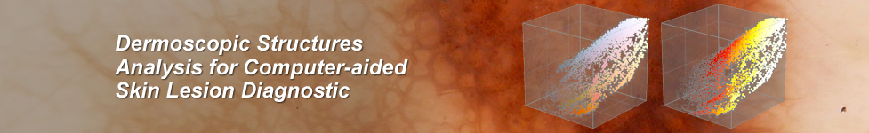
Diagnosis of skin nevi involves visual observation and finding additional information, such as growth rate, the presence of other signs, family history.
Common in area of diagnosis is video - dermatoscope, which is a digital camera with additional lens system, equipped with halogen illuminator and magnifying lenses (4-100X). Analysis of taken image is made on a computer monitor. Purpose of this work is to analyze the characteristics of wavelet images taken only in the light of white.
Clinical examination and an interview based on non-invasive, visual examination of lesion require a lot of experience. But even clinical experience is not sufficient to identify with reasonable statistics melanomas in the early phase, when the characteristics are not yet visible. The only sure way is to perform biopsy, but this method is invasive and cannot be applied to a large number of lesions, or when there are other contraindications. Furthermore histopathology requires specialization in field of skin tissue analysis, otherwise often "redundant" or "safe" interpretation is made and lesion is diagnosed as melanoma.
Because accurate diagnosis and prognosis of melanoma is based on an analysis of its vertical profile (growth into the dermis), and the histopathological examination is in some cases may be impossible other methods of lesion surface imaging may "extract" the vertical profile of the skin. For this reason, methods supporting dermatoscopic image recognition are of great interest because of early non-invasive diagnosis of pigmented moles is a factor potentially saving lives.
The use of machine learning techniques in medical diagnosis support systems is not a new approach. Computational intelligence methods often find practical application in medicine and have proven their usefulness. Therefore, there are compelling reasons to believe that the computer analysis of dermatoscopic images and patient history, can improve the sensitivity and / or specificity for early diagnosis of potentially cancerous moles. This is confirmed by the results of research conducted by the authors of the proposal. The development of such methods and diagnostics system based on them would be a great help for the doctor, but also for the patient who in most cases in the present state of knowledge is condemned to invasive therapy.
The research project is important practical application in the early diagnosis of malignant melanoma.
Project leader: Prof. Maciej Ogorzałek
Project title: Application of Computational Intelligence Techniques in Analysis of Images for Computer-Assisted Melanoma Diagnosis
Project value: 295 000 PLN
Project duration: 36 months
Project start: 16 April 2010
Project ends: 15 April 2013
For details see Melanoma Computer-aided Diagnostics project.
Grant related publications
- K. Przystalski, L. Nowak, M. Ogorzalek, G. Surowka (2012). Applications of Neural Networks in Semantic Analysis of Skin Cancer Images, Human – Computer Systems Interaction: Backgrounds and Applications 2 Series, Advances in Intelligent and Soft Computing, Vol. 99, pp. 111-124, Hippe, Zdzisław S.; Kulikowski, Juliusz L.; Mroczek, Teresa (Eds.), ISBN 978-3-642-23171-1
- M.J. Ogorzałek, L. Nowak, G. Surówka, A. Alekseenko (2011). Modern Techniques for Computer-Aided Melanoma Diagnosis, Melanoma in the Clinic - Diagnosis, Management and Complications of Malignancy, Prof. Mandi Murph (Ed.), ISBN: 978-953-307-571-6, InTech, Available from: http://www.intechopen.com/articles/show/title/modern-techniques-for-computer-aided-melanoma-diagnosis
- K. Przystalski, M. Popik, M.J. Ogorzałek, L. Nowak (2011). Improved Melanoma Diagnosis Support System Based on Fractal Analysis of Images, ISORA 2011 August 28-31, 2011,Dunhuang, China, ORSC & APORC, pp. 203–210
- W. Piatkowska, J. Martyna, L. Nowak, K. Przystalski (2011). A Decision Support System Based on the Semantic Analysis of Melanoma Images Using Multi-elitist PSO and SVM, MLDM 2011, Lecture Notes in Computer Science, pp. 362-374
- K. Przystalski, L. Nowak, M.J. Ogorzałek, G. Surówka (2010). Semantic Analysis of Skin Lesions Using Radial Basis Function Neural Networks, Human System Interactions, pp. 128 – 132, ISBN: 978-1-4244-7560-5
- K. Przystalski, L. Nowak, M. Ogorzałek, G. Surówka (2010). Decision Support System for Skin Cancer Diagnosis, ISORA 2010, Lecture Notes in Operations Research 12, pp. 406–413, ISBN: 978-7-5100-2406-1/O819, APORC, Word Publishing Corporation
- W. Piątkowska, J. Martyna, L. Nowak, K. Przystalski (2010). A Decision Support System Based on Semantic Analysis of Melanoma Images Using SVM, MICAI 2010
- Grzegorz Surówka (2010), Symbolic learning supporting Early diagnosis of melanoma", IEEE EMBC 2010 Buenos Aires


